
What to do if the skin of a dog is phytosomiasis and pigment disorder?
How to deal with phytoniasis and pigment disorder in dogs:
---------------------------------------------------------------------------------------------------------------------------------------------------------------------------------------------------------------------------------------------------------------------------------------------------------------------------------------------------------------------------------------------------------------------------------------------------------------------------------------------------
1. Microsporidium canis exist in the canine body for a long time and only produces mild inflammation. About 50% of the phytopathy in canines is caused by this bacteria.
2. Gypsum-like microsporidium is a soil-philic fungus. The warm climate occasionally causes phytopathy in dogs, but this inflammatory response and infection are self-limiting.
3. Microsporeus mainly causes secondary phytosomiasis in dogs, and mice are the main carriers.
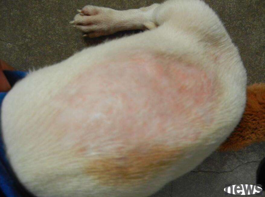
Dermatitis is mainly caused by mycelium invasion of hair columns, hair follicles and stratum corneum, causing hair loss and dander. It often occurs in the head, feet and legs, and is prone to adult dogs in small dogs.
【Diagnosis points】
Diagnosis
①Take hair or skin shaves. Observed under Wood's lamp, the dog hair infected with Microsporidium canis showed green fluorescence.
②Digestible potassium cyanoxide hot solution and scrape the extract, and place it under a microscope to see if there are any fungal seeds.
③ The disease material was inoculated in Shabuca's culture medium and cultured for 1-2 weeks at room temperature. The colonies of the Microsporidium canis were yellow basal surface and were fine curly hair. The Microsporidium was brown-based and the ground was covered with fine particles. The gypsum-shaped microsporidium colonies were yellow leather-like; the black or blue colonies were contaminated fungi. Adding phenol red to Shabuca's culture medium can turn red colonies red can be preliminarily diagnosed as dermatitis.
Treatment The preferred drug is greyfulvin, with a daily dose of 15mg/kg, and feed high-fat foods to promote intestinal absorption. Local antibacterial preparations such as clotrimazole can be removed locally. Local infection of demodex is common on the face and legs. Most diseased dogs are chronic. Some dogs develop into deep tissue suppuration. There are mainly two changes in skin inflammation of the demodex disease:
① Local hair is sparse or completely dehaired, the skin is thickened, the dandruff increases and becomes dark, and the swollen hair follicles can squeeze out waxy-like sebum.
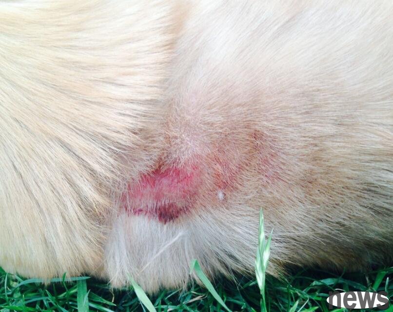
② Destroy hair follicles and reach the dermis, causing skin ulcers or swelling of the flesh, secondary bacterial infection, which becomes pyoderma.
【Diagnostic Points】
Treatment Treatment of large-area demodex infection is relatively difficult. During treatment, first cut off the inflammatory area and surrounding hair, clean it with alcohol, etc., and take a bath with sulfur soap or shampoo to remove the surface skin. Locally use low-toxic, efficient and easy-to-permeable drugs, such as lithosulfur mixture, 0.5% DDT, zombie, etc., which can be cured. Some people abroad have introduced that using O.75% alcohol otamantein is more effective. If a dog is infected with bacteria, it should be considered to use antibiotics locally or completely to treat
2. Pigment disorder
Pigment disorder, and there are often the following two types in clinical practice.
(I) Black acanthosis
This is a skin disease in dogs characterized by papillary hyperplasia, excessive keratosis of the epidermis and increased pigmentation. It is more common in German shepherds and beagles. The cause is related to endocrine disorders, especially thyroid disorders. Clinically, diseased dogs often occur in the face and ventral parts and are symmetrical. The affected part is covered with hair, the skin gradually becomes thicker and rough, the dandruff increases, and the color gradually becomes darker.
Chronic dermatitis in some dogs, such as demodex, seborrheic dermatitis, and vitamin A deficiency, can also appear similar acanthosis. Pay attention to distinguishing it during treatment.
【Diagnostic Points】
For treatment, you can take orally thyroxine preparations, such as triiodothyroxine, at 1.0mg/Kg per day, and the course of treatment is 1 month. Severe sick dogs can use corticosteroids, thyroxine and iodine preparations, etc., and local treatment can be used to wash the dog's body with sulfur soap, and then apply corticosteroid drugs.